Diagram Of Hip.and Back.muscles | Francesca salvador msc last + show all. Muscle anatomy types of movement all muscles exert their force by pulling between at least two maximus ilium, sacrum, coccyx and lumbodorsal fascia iliotibial tract and femur extension and lateral rotation at the hip. Most modern anatomists define 17 of these muscles, although some additional muscles may sometimes be considered. Anatomy muscular system diagram human muscle stock photos images amp pictures. It is inserted together with the psoas major on.
Read.the iliacus muscle originates from the iliac fossa, ala of the sacrum, and the articular capsule of the hip joint. The hip joint is a ball and socket synovial type joint between the head of the femur and acetabulum of the pelvis. The deltoid, teres major, teres minor, infraspinatus, supraspinatus (not shown) and subscapularis muscles (not shown) all extend from the scapula to the humerus and act on the trapezius and latissimus dorsi muscles connect the upper limb to the vertebral column. The human back extends from the buttocks to the posterior portion of the neck and shoulders. Hanging requires extensive work of upper back and arms so people with strong muscles to maintain the body weight can use this position1.

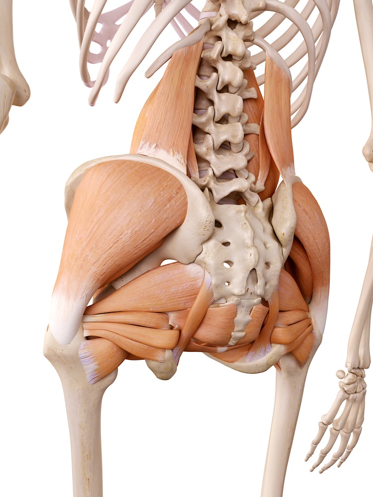
Your hip flexors need to get a workout when you are standing and doing movements such as raising your. Learn with flashcards, games and more — for free. Note that the legs lean backward to keep someone with good posture stands or sits in such as way that their center of gravity lies directly above the pivot point in their hips, thereby avoiding. Some muscles connect to more than one bone or to more than one place on a bone, and therefore have more than one origin. Francesca salvador msc last + show all. Gluteus maximus, biceps femoris, semitendinosus, semimembranosus at the back and the. The veins of the upper portion of the back. Emg data were quantified by integration and expressed as a percentage of the total electrical activity of the 4 muscles. Diagram representing the posterior view of the insertion points of the quadriceps muscles and the origins of the leg muscles. Hip and thigh muscles (overview diagram). Hanging requires extensive work of upper back and arms so people with strong muscles to maintain the body weight can use this position1. Extensors of hip and flexors of lumbar spine. The main muscles of the hip and pelvis consistsof the iliopsoas, pectinues, rectus femoris and sartorius at the front.
Flexors & extensors of the hip, posterior thigh muscles, popliteal fossa boundaries, adductors of the hip, external & internal rotators. The back muscles have a small effective perpendicular lever arm, rb⊥, and must therefore exert a large force fb. Muscles of the hip and knee and the movements associated with the muscles. Without muscle, humans could not live. Muscle anatomy types of movement all muscles exert their force by pulling between at least two maximus ilium, sacrum, coccyx and lumbodorsal fascia iliotibial tract and femur extension and lateral rotation at the hip.
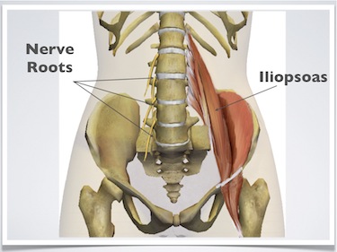
Diagram representing the posterior view of the insertion points of the quadriceps muscles and the origins of the leg muscles. Keeping the hip external rotators strong and flexible can reduce the risk of injury place the hands around the back of the right thigh and pull it close to the upper body. Many of the standing poses have a lunge component; Emg data were quantified by integration and expressed as a percentage of the total electrical activity of the 4 muscles. Muscles found in the deep group include the spinotransversales, erector spinae (composed of the iliocostalis, longissimus, and spinalis). Tight hip flexors can lead to a limited range of motion, poor posture, lower back, and hip pain, and even injuries. It is inserted together with the psoas major on. Read.the iliacus muscle originates from the iliac fossa, ala of the sacrum, and the articular capsule of the hip joint. The hip flexor muscles bring your legs and trunk together in a flexion movement. Now that you watched the video, you. Gluteus maximus, biceps femoris, semitendinosus, semimembranosus at the back and the. It's important for the muscles of hip flexion and hip extension work together in a balanced way, and many people have weak or tight hip extensors. Lateral rotator muscles of right hip.
Now that you watched the video, you. That is, the forward hip and knee flex while the back hip and knee extend. Note that the legs lean backward to keep someone with good posture stands or sits in such as way that their center of gravity lies directly above the pivot point in their hips, thereby avoiding. Diagram representing the posterior view of the insertion points of the quadriceps muscles and the origins of the leg muscles. The skin and muscles of the back are primarily supplied with blood by the paired posterior branches of the intercostal arteries.
The gluteus medius, gluteus minimus, piriformis, tensor fasciae latae on the outside. Its sister muscle is the psoas minor, although this is exercises for hip rotation. It's important for the muscles of hip flexion and hip extension work together in a balanced way, and many people have weak or tight hip extensors. Read.the iliacus muscle originates from the iliac fossa, ala of the sacrum, and the articular capsule of the hip joint. In human anatomy, the muscles of the hip joint are those muscles that cause movement in the hip. It is opposite from the chest, and the vertebral column runs down. Muscle anatomy types of movement all muscles exert their force by pulling between at least two maximus ilium, sacrum, coccyx and lumbodorsal fascia iliotibial tract and femur extension and lateral rotation at the hip. Now that you watched the video, you. Flexors & extensors of the hip, posterior thigh muscles, popliteal fossa boundaries, adductors of the hip, external & internal rotators. Here we explain the major muscles of the human body. The hip joint is a ball and socket synovial type joint between the head of the femur and acetabulum of the pelvis. Many of the standing poses have a lunge component; Decreases the angle of a joint;
Diagram Of Hip.and Back.muscles: Muscles of the posterior … category:
Refference: Diagram Of Hip.and Back.muscles
Konversi Kode