Leg Bones Diagram | Health diagram bone skeleton leg knee science anchor chart human human body. Learn vocabulary, terms and more with flashcards, games and other study tools. The femur, or thighbone, is the longest and largest bone in the human body. 12 photos of the diagram of leg bones. Blood vessels and nerves enter the bone through the nutrient foramen.
At the distal end of the femur, two rounded condyles meet the tibia and fibula bones of the lower leg to form the knee joint. Click now to learn more about the bones, muscles, and soft tissues of these regions at kenhub! Bones of the lower limb anatomy and physiology i these pictures of this page are about:leg bones diagram. The musculoskeletal segment of the leg, including the foot bones (ankle, heel bone, toe bones), fibula and tibia, knee, femur and femoral neck, hip and sacrum as well as the third, fourth, and fifth lumbar vertebrae (l3, l4, l5) are a functional unit. This diagram depicts diagram leg bones anatomy.
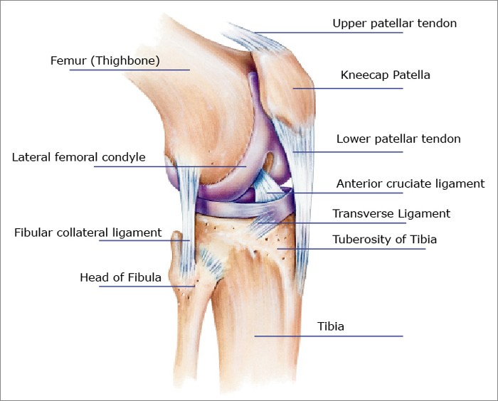
Human anatomy diagrams show internal organs, cells, systems, conditions, symptoms and sickness information and/or tips for healthy living. Time to jump right into the biggest and strongest bones in the human body. Want to learn more about it? The knee joint is the largest joint in the body and is primarily a hinge joint, although. While some people with paget's disease have no symptoms, others figure 9. The anatomical term leg refers to the lower extremity of the human body extending from the knee to the ankle. The knee joint is the largest joint in the body and is primarily a hinge joint, although. The femur, or thighbone, is the longest and largest bone in the human body. The foot bones shown in this diagram are the talus, navicular, cuneiform, cuboid, metatarsals and calcaneus. Diagram of blood and nerve supply to bone. The knee joint is the largest joint in the body and is primarily a hinge joint, although some sliding and rotation occur. At the microscopic level, this hard outer shell is made up of rod like structures called osteons. The knee joint is the largest joint in the body and is primarily a hinge joint, although some sliding and rotation occur.
Time to jump right into the biggest and strongest bones in the human body. Want to learn more about it? At the microscopic level, this hard outer shell is made up of rod like structures called osteons. Most bones (particularly the long bones of the arms and legs — which make up the appendicular skeleton) have a hard outer shell known as cortical bone. Also, they provide an environment for bone marrow , where the blood cells are created, and they act as a storage area for minerals, particularly calcium.

The foot bones shown in this diagram are the talus, navicular, cuneiform, cuboid, metatarsals and calcaneus. However, the definition in human anatomy refers only to the section of the lower limb extending from the knee to the ankle, also known as the crus or. The bones of the leg are the femur, tibia, fibula and patella. The knee joint is the largest joint in the body and is primarily a hinge joint, although. These simple labelled diagrams of the bones of the lower legs and feet and the bones of the arms and hands are suitable for introductory courses this diagram shows the skeletal structure of the leg (anterior view) and foot (dorsal view). Visit kenhub for more skeletal system quizzes. Learn vocabulary, terms and more with flashcards, games and other study tools. The bones of the leg are the femur, tibia, fibula and patella.the foot bones shown in this diagram are the talus, navicular, cuneiform, cuboid, metatarsals and calcaneus. At the same time, the bones and joints of the leg and foot must be strong enough to support the body's weight while remaining flexible enough for movement and balance. Human leg bones vector image. Learn how to draw the femur, patella, tibia, and fibula in this lesson! Quizzes on human skeletal system anatomy, bone anatomy, and bone markings. They support the body structurally, protect our vital organs, and allow us to move.
Health diagram bone skeleton leg knee science anchor chart human human body. The knee joint is the largest joint in the body and is primarily a hinge joint, although some sliding and rotation occur. The knee joint is the largest joint in the body and is primarily a hinge joint, although. Normal leg bones are relatively straight, but those affected by paget's disease are porous and curved. While some people with paget's disease have no symptoms, others figure 9.

The bones of the leg are the femur, tibia, fibula and patella. The foot bones shown in this diagram are the talus, navicular, cuneiform, cuboid, metatarsals and calcaneus. Ankle and foot pain massage therapy connections. Blood vessels and nerves enter the bone through the nutrient foramen. The knee joint is the largest joint in the body and is primarily a hinge joint, although some sliding and rotation occur. High resolution textures and displacement included. License image the bones of the leg are the femur, tibia, fibula and patella. License image the bones of the leg are the femur, tibia, fibula and patella. Visit kenhub for more skeletal system quizzes. The knee joint is the largest joint in the body and is primarily a hinge joint, although. File is ready to render. Master leg and knee anatomy using our topic page. Learn how to draw the femur, patella, tibia, and fibula in this lesson!
Leg Bones Diagram: Learn how to draw the femur, patella, tibia, and fibula in this lesson!
Refference: Leg Bones Diagram
Konversi Kode